Hydrocephalus
Key Points
- Programmable shunts are used routinely. This has significantly reduced the rate of shunt failures
- Infection Rate is < 1.8%
- Referral Center for the complex hydrocephalus patients
- Quick Scan MRI: No sedation, No Radiation and is routinely used for brain imaging
What is Hydrocephalus?
The brain and the spinal cord are bathed in a fluid called Cerebral Spinal Fluid (CSF). CSF is distributed throughout the brain and spinal cord via a complex plumbing system. There are many large parts to this plumbing system. Fluid is produced in the ventricles (third, fourth and lateral). We liken it to a faucet that can never be turned off. The fluid then passes thru small channels, like sink holes in the brain, the cerebral aqueduct, and the interventricular foramen. CSF eventually gets reabsorbed by microscopic small holes called arachnoid granulations on the surface of the brain. The arachnoid granulations are like pipes that carry the fluid from the sink holes as they leave the house.
Anytime there is a blockage in one of the channels of the brain or the arachnoid granulations, the plumbing system can get backed up. That backup can cause increased pressure in the brain because CSF is still produced in spite of the blockage. This condition is called hydrocephalus. Hydrocephalus is dangerous because it damages the brain, and ultimately can be fatal.
Hydrocephalus Treatment
Hydrocephalus is treated surgically to bypass this blockage. Commonly, a shunt, which is a flexible tube, is used to bypass the blockage. One end of the tube is inserted into a brain ventricle. The other end of the tube is usually placed in the abdominal cavity and the excess CSF is absorbed there. The shunt includes a valve mechanism, which controls the flow of CSF through the tubing. A shunt can sometimes become clogged or malfunction and another surgery may be required to fix it. To reduce the chance of this blockage, we routinely utilize shunts that can be programmed to adjust the CSF flow. When the sink holes are blocked, but the pipes are open, a bypass can be created at the base of the brain with an operation called endoscopic third ventriculostomy. As long as this bypass stays open, a medical devise like the shunt would not be needed.
Children with hydrocephalus require frequent imaging of the brain. We now routinely use " quick scan" to avoid the radiation of a CT and the sedation required for a regular MRI.
Shunt Infections
Infection is one of the complications of shunt operations. Once there is an infection in the tubing, additional surgery is needed to replace the shunt. Antibiotics alone will not reliably kill all the bacteria in the shunt tube. We take great care in our surgical protocol to reduce our infection rate to less than 1%. The national average is 4-10%. The following illustrates the key features of our protocol for preventing shunt infections.
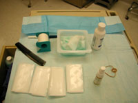
Key features of our protocol:
- Minimal hair removal. We only remove the hair necessary to perform the surgery.
-
Our surgical team cleanses the skin with alcohol, shampoo with chorohedine, prep with betadine and duraprep. (trade name)

-
The child's skin is protected and covered with antibacterial adhesive during surgery.
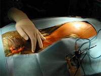
-
During the surgery, only minimal traffic is allowed in and out of the operating room.

- IV, topical and CSF antibiotics are used post operatively to prevent infection.